We tested whether the endoderm cells require the rest of the embryo to go through invagination. We used a tiny micropipette needle to cut the embryo in half and separate the vegetal half of the blastula from the animal half, and then filmed it to see how the endoderm would behave compared to an intact embryo [new window, 520x580 pixels, 3.8 MB]. A vegetal hemisphere was isolated at the 64 cell stage. A time-lapse movie was taken of this plate through the time when normal invagination occurs. The endoderm cells appeared to invaginate while the surrounding cells curled upward. This suggests that the endoderm can invaginate as part of the vegetal hemisphere only. Therefore, the animal hemisphere is not necessary for invagination to occur. You will see in this video that these cells are able to invaginate without the animal pole cells adhered to the embryo and that the cells remember which is their apical surface and which is the basal surface because they dive in the same direction that endoderm cells do in an intact embryo.
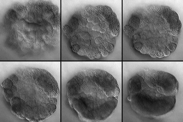
Frames from a time-lapse movie of an isolated Boltenia vegetal plate. Click the picture to see the reduced-resolution movie [new window, 560x620 pixels, 4.2 MB] or view the full-size version [new window, 700x750 pixels, 19.8 MB]. Posterior is toward the top of the picture, the endoderm cells are in the center of the isolate. At first, the half-embryo is a dome with the endoderm in focus (top left); endoderm cells divide and the dome flattens (top middle and right); the endoderm cells go out of focus slightly as they begin to invaginate (bottom left) but then grow taller as they wrap the surrounding cells over them (bottom middle and right). The dark cells that are out of focus are the two patches of muscle precursors and (top left and right in each image) and the arc of neural plate and notochord cells (bottom of the image).
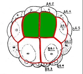
Having observed that the vegetal half embryo does not require the presence of animal pole cells in order to invaginate, we became interested in observing the behavior of isolated endoderm cells. Using micromanipulations, we isolated the A6.1 cells at the 32 cell stage.
 |
 |
This is the vegetal hemisphere of a 32 cell stage embryo. The A6.1 cells are highlighted in green. |
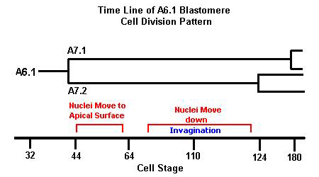
The cell division pattern and nuclear migration of A6.1 at different cell stages of an intact Boltenia embryo |
In a whole embryo, the A6.1 cell cleaves at the 44 cell stage into A7.1 and A7.2. Both of these cells, in intact embryos, participate in the earliest steps of invagination, and all their descendants form endoderm. The next time A7.1 and A7.2 divide is after endoderm invagination is complete. The nuclei in A7.1 and A7.2 move to the apical surface after the 44 cell stage and they dive down into the cell as invagination starts.
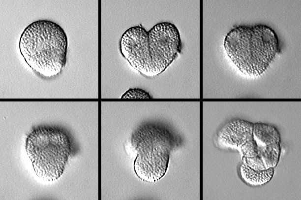
Frames from a time-lapse movie of an isolated A6.1 cell. Click the picture to see the movie [new window, 400x500 pixels, 2.3 MB]. Isolated A6.1 cells round up and divide (top left and middle); nuclei move close to one surface (top right), then migrate together (bottom left) before descending as the cell moves downward (bottom middle). These isolated cells' descendants survived >12 hours and made an epithelium with a lumen, like endoderm cells would in the whole embryo.
The A6.1 cell divides, the nuclei rise to the top of the A7.1 and A7.2 cells, and then migrate down. Notice how the nuclei move close to each other before they begin moving down. This isolated cell displays behavior reminiscent of invagination of endoderm cells in the whole embryo as described in the A6.1 cell division time line. This suggests that the endoderm cells are capable of invaginating autonomously and the forces that drive invagination must be intrinsic to the endoderm cells. We observed this behavior in multiple A6.1 cells isolated at the same time. The nuclei of all the isolated cells migrate downward at the same time, implying that a coordinated, cell-autonomous process of cell shape change drives invagination.
