3-D reconstructions ( color code and general features as in Fig 3.) of prophase 10 (rows 1 and 2) and metaphase 10 (rows 3 and 4), both from apical vantages. The prophase density of microtubules is higher than at any other phase, but few microtubules connect back to centrosomes. Microtubule geometry is extremely complex in prophase compared to early interphase 3 minutes earlier (in Fig. 3), and compared to metaphase 2 minutes later, when microtubules are scarce, except for spindle microtubules. In prophase, F-actin forms figures of eight linked rings above some nuclei, and ellipsoidal rings above others, always with the centrosomes at the foci.
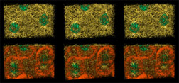
The prophase 10 reconstruction is of a 42 micron wide by 28 micron high by 21 micron deep volume of the syncytial blastoderm of a Drosophila embryo fixed in prophase of mitotic cycle 10 (at a time corresponding to two minutes following the early interphase 10 situation the previous reconstructions show). The density of microtubules has increased dramatically. Most are no longer traceable back to the centrosomes which presumably nucleated their polymerization. The F-actin caps present 2 minutes ago in early interphase have become hollow rings, surrounding each centrosome, which join to make figure of 8 shapes. The chromatin has started to condense.
A combined view of Fig. 4 may be viewed in two larger sizes, opening in new windows sized as indicated, the large version [840 x 600 pixels, 112K] or the largest version [1530 x 800 pixels, 0.9MB].
QuickTime version 3 or higher is required for these movies. Visit the apple website to download the latest, free version of QuickTime.
|
3D virtual object movies (interactive) |
tiny [15 MB] |
half size [34 MB] |
full size [110 MB] |
|---|---|---|---|
|
movies (passive viewing) |
half size [25 MB] |
full size [640 X 480 pixel, 218 MB] |
|
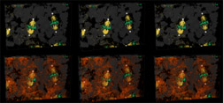
The metaphase 10 reconstruction shows a 42 micron wide by 28 micron high by 19 micron deep volume of the syncytial blastoderm of a Drosophila embryo fixed in metaphase of mitotic cycle 10. Note how few microtubules exist at metaphase compared to the number that exist in the preceding prophase and the following anaphase.
A combined view of Fig. 4 may be viewed in two larger sizes, opening in new windows sized as indicated, the large version [840 x 600 pixels, 112K] or the largest version [1530 x 800 pixels, 0.9MB].
QuickTime version 3 or higher is required for these movies.
|
3D virtual object movies (interactive) |
tiny [10 MB] |
half size [18 MB] |
full size [60 MB] |
|---|---|---|---|
|
movies (passive viewing) |
half size [19 MB] |
full size [640 X 480 pixels, 183 MB] |
|
pseudo-cleavage and buds project • back
