
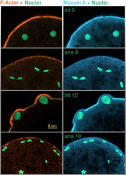
Cross sectional LSCM images of the Drosophila embryo anterior showing F-actin (red), myosin II (blue) and nuclei (green) during cycle 9 interphase (top row), anaphase 9 (row 2), cycle 10 interphase (row 3) and cycle 10 anaphase (bottom row). F-actin is visualized with Bodipy FL phallicidin, nuclei with propidium iodide, and myosin II by indirect immunofluorescence using a polyclonal antibody we prepared against the myosin tail (see Materials and Methods in the complete text [MS Word RTF 196K]).
This image may be viewed in two larger sizes, opening in new windows sized as indicated, the large version [840 x 600 pixels, 68K] or the largest version [1600 x 800 pixels, 1.4MB].
The time-lapse movie called Anterior Buds (three size options) shows the time course of periodic bud eruption and collapse. Cytoplasmic buds forming as migrating nuclei arrive at embryo's anterior end in cycle 10. Movie ends in cycle 14 with cellularization.
QuickTime version 3 or higher is required for these movies. Visit the apple website to download the latest, free version of QuickTime.
|
Anterior Buds time-lapse movies (passive viewing) |
tiny [13 MB] |
half size [63 MB] |
full size [690 X 680 pixels, 188 MB] |
|||
|---|---|---|---|---|---|---|
pseudo-cleavage and buds project • back