Research Associate
atdawes@u.washington.edu
All multicellular organisms, including humans, begin life as a single fertilised cell. During development, the embryo must have a mechanism to specify cell fate to properly differentiate cells in the mature organism. The nematode worm C elegans initiates cell fate specification in the fertilised embryo by segregating specific molecules to opposite ends of the cell. This polarized segregation is maintained through first division, resulting in biochemically distinct daughter cells. Stable intracellular polarization is a fundamental biological process, seen not just in C elegans embryos, and is crucial for wound healing, immune surveillance and embryonic development in many organisms. While a number of experimental biologists have been studying polarization in many different biological contexts, the exact mechanism is not well understood. However, these experiments have uncovered many similarities among the key signalling molecules involved in polarization in these different contexts. As a post doctoral fellow, I propose to study the establishment and maintenance of polarity in the early C elegans embryo using both theoretical and experimental techniques in an effort to increase our understanding of this important biological process.

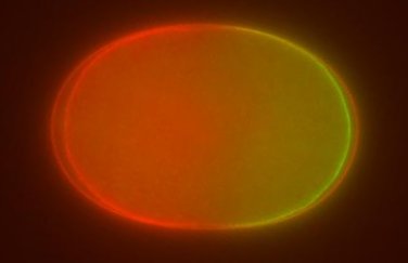
Image of single-celled embryo at centration showing spatial segregation of the PAR proteins. PAR-6 is tagged with red fluorescent protein (RFP) and PAR-2 is tagged with green fluorescent protein (GFP). Anterior is to the left, posterior is to the right. Image credit: Adriana Dawes
Most experiments to study polarization in the C elegans embryo have focussed on the PAR proteins, a class of proteins that are crucial for proper polarization of the embryo1. The PAR proteins include PAR-3, PAR-6 and aPKC which localize to the anterior end of the embryo, and PAR-1 and PAR-2 that localize to the posterior. These proteins interact with each other and occupy distinct spatial domains in the embryo, as shown above. I have begun to study the PAR proteins by developing continuum models of coupled ordinary and partial differential equations based on experimentally determined interactions. The models suggest that stable polarization depends not only on mutually inhibitory interactions between the PAR proteins, but also on the ability of one of the PAR proteins to bind to itself and form oligomers, allowing the system of equations to admit bistable solutions and challenging the current belief that only mutual inhibitory interactions are required to maintain polarity. The model results also suggest a number of experimentally testable hypotheses. Using RNA interference to modulate the expression of certain proteins, I can image live C elegans embryos that have PAR proteins tagged with fluorescent markers to directly test the assumptions and consequences of the model. Early results are consistent with the model predictions although further work is required to rule out the possibility that the spatial patterning is caused by a reaction-diffusion Turing type mechanism.
More recent experimental work has determined that mechanical processes, particularly remodelling of the actin cortex (a structure that provides structural stability to the cell), are required for proper polarization2. I am using software developed by Ed Munro to explore cortical dynamics and how this regulates and is regulated by protein networks such as the PAR proteins. This approach will generate predictions that can be tested using appropriate experiments, using a variety of imaging techniques and working with both live and fixed worm embryos. The combination of theoretical, computational and experimental techniques will help us develop more complete and accurate models of polarization in the C elegans embryo, which could provide insight into other instances of cell polarization.